- SHOWA Medical University Hospital
- Surgery for Hemifacial Spasm and Trigeminal Neuralgia
- Japanese
- English
Hemifacial Spasm and Trigeminal Neuralgia
Hemifacial Spasm and Trigeminal Neuralgia
Microvascular Decompression Surgery
Less invasive Microvascular Decompression Technique Performed in the Supine Position Without Brain RetractorsDr. Shimizu specializes in delicate microsurgical procedures, including keyhole surgery and skull base tumor surgery, and has been featured in several respected medical information resources, including:
- Doctor's Guide (Jiji Press Publishing) http://dr-guide.net/www/
- Neurosurgical Specialists Directory http://doctor-cancer.com/cranialnerves-dr.html
- Brain Disease Web (featured among leading neurosurgeons) Listed among renowned neurosurgeons
In addition, the Comprehensive Center for Trigeminal Neuralgia and Hemifacial Spasm has been established at Showa University Hospital to provide specialized multidisciplinary care for patients with these conditions.
Media Appearances
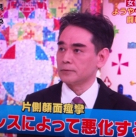
Dr. Shimizu was featured as the treating physician and surgeon for a well-known actress who successfully recovered from hemifacial spasm surgery. His work was highlighted on Fuji Television's FNN Super News in 2010 and TBS's Hiruobi!in 2011. He also appeared on the television program Takeshi's Health Entertainment! Family Doctor in 2012.A Unique and Established Microvascular Decompression Technique
Since 1985, Dr. Shimizu has performed microvascular decompression (MVD), the definitive surgical treatment for hemifacial spasm and trigeminal neuralgia, using a technique specifically designed to minimize the burden on patients.This procedure is performed with the patient in the supine position (lying on the back), which is considered the most natural body position. The technique has been published in the medical literature and established as a distinctive surgical approach.
Unlike conventional MVD procedures, this method:
- Utilizes the natural supine position throughout surgery
- Avoids the use of brain retractors, which are commonly employed to retract neural tissue during the procedure
By eliminating the need for brain retractors and maintaining patients in a natural position, this approach aims to provide a gentler and less invasive surgical experience.
Advanced Microsurgical Expertise in Skull Base Surgery
This technique has also been successfully applied to acoustic neuroma (vestibular schwannoma) surgery and other skull base tumor procedures.Drawing upon extensive experience in the management of complex skull base disorders, Dr. Shimizu is recognized for performing highly delicate microsurgical operations in regions where critical nerves and blood vessels are densely concentrated. His surgical philosophy emphasizes both safety and effectiveness while striving to minimize patient burden.
Hemifacial Spasm
The typical symptoms of hemifacial spasm usually begin with intermittent twitching of one eyelid. As the condition progresses, patients may develop facial tightness in the cheek and involuntary pulling of the corner of the mouth on the same side. In more advanced cases, the eyelid may remain closed for prolonged periods, and the entire affected side of the face may become markedly distorted by continuous spasms.These movements occur completely involuntarily and are unrelated to the patient's intentions. Symptoms are often aggravated by stress, fatigue, anxiety, or emotional tension.
Hemifacial spasm remains relatively unfamiliar to the general public, and awareness is still limited even among healthcare professionals. As a result, some patients experience considerable difficulty obtaining an accurate diagnosis despite seeking medical attention.
In the vast majority of cases, the underlying cause is compression of the facial nerve, which controls the muscles of facial expression, by a blood vessel inside the skull. This compression typically occurs near the point where the facial nerve exits the brainstem, an area known as the root exit zone (REZ). Constant pulsatile irritation from the offending vessel places the nerve in a state of chronic hyperexcitability, resulting in involuntary facial contractions.
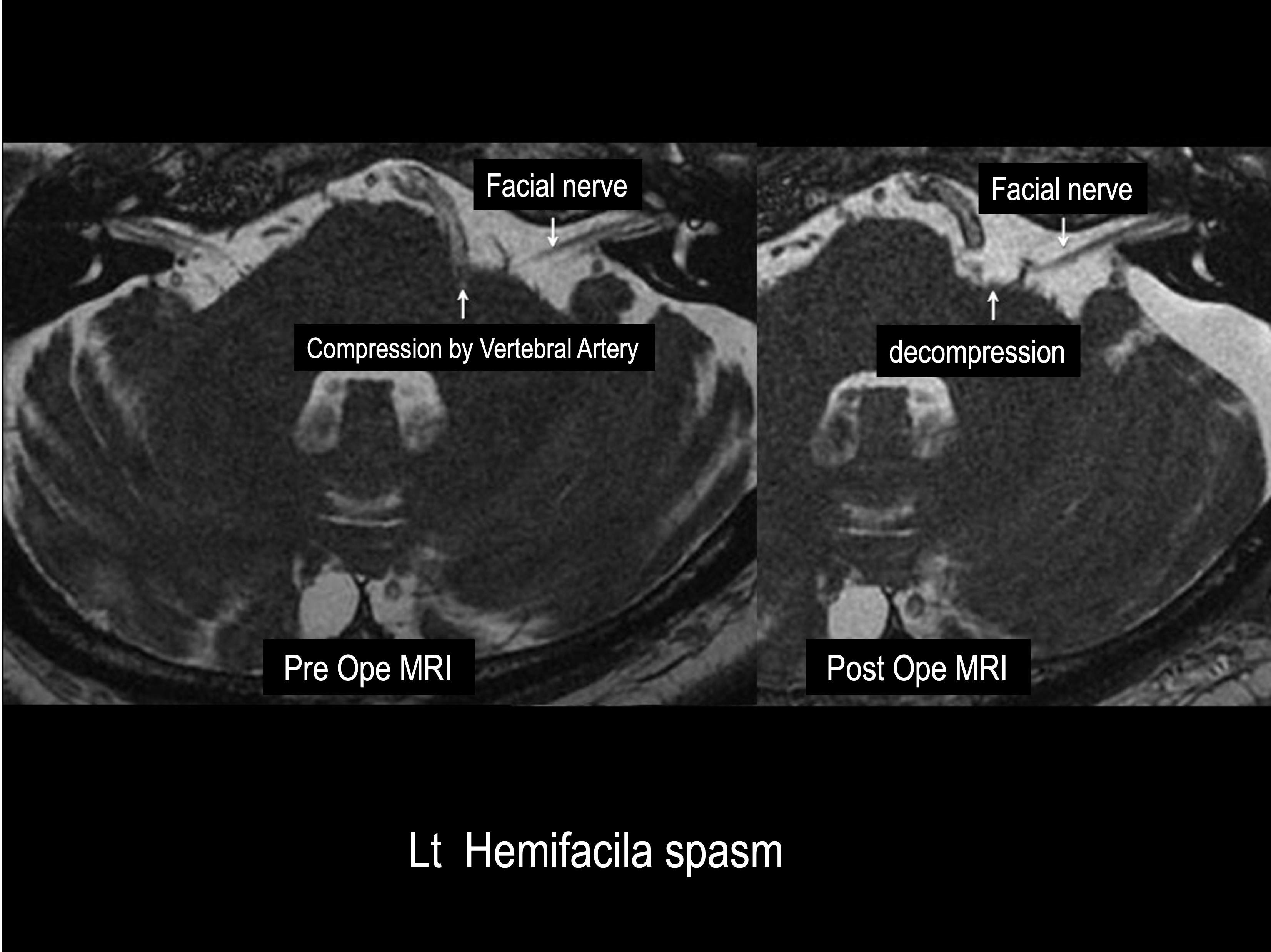
To achieve definitive treatment, the underlying cause of the nerve compression must be addressed surgically. This is accomplished by repositioning the offending blood vessel and relieving the pressure exerted on the facial nerve near the REZ.
This procedure is known as microvascular decompression (MVD), or Jannetta's procedure. During the operation, a small opening approximately the size of a coin is created in the skull behind the ear, and the surgery is performed under an operating microscope.
Microvascular decompression is often referred to as keyhole surgery because it is performed through a very small craniotomy. Despite the limited opening, the surgeon must work within the highly complex region of the brainstem, where numerous critical nerves and blood vessels are concentrated. As such, MVD is a delicate microsurgical procedure that requires advanced technical expertise and extensive surgical experience.
Trigeminal Neuralgia
Trigeminal neuralgia is characterized by sudden, severe facial pain. Patients may experience excruciating pain radiating from the jaw to the cheek while speaking, chewing, or brushing their teeth. Others develop intense, localized pain in the forehead, around the eye, or around the mouth when exposed to cold wind or even light touch to the face.Because the pain can closely resemble that of a toothache, many patients initially consult a dentist before trigeminal neuralgia is correctly diagnosed. At our institution, close collaboration between the medical and dental faculties enables comprehensive evaluation and treatment of patients with facial pain disorders.
Although trigeminal neuralgia can have various causes, one of the most common mechanisms is similar to that of hemifacial spasm. The trigeminal nerve, which transmits sensory information from the face, may be compressed by a blood vessel near its exit from the brainstem. Continuous pulsatile compression of the nerve can lead to episodes of severe facial pain.
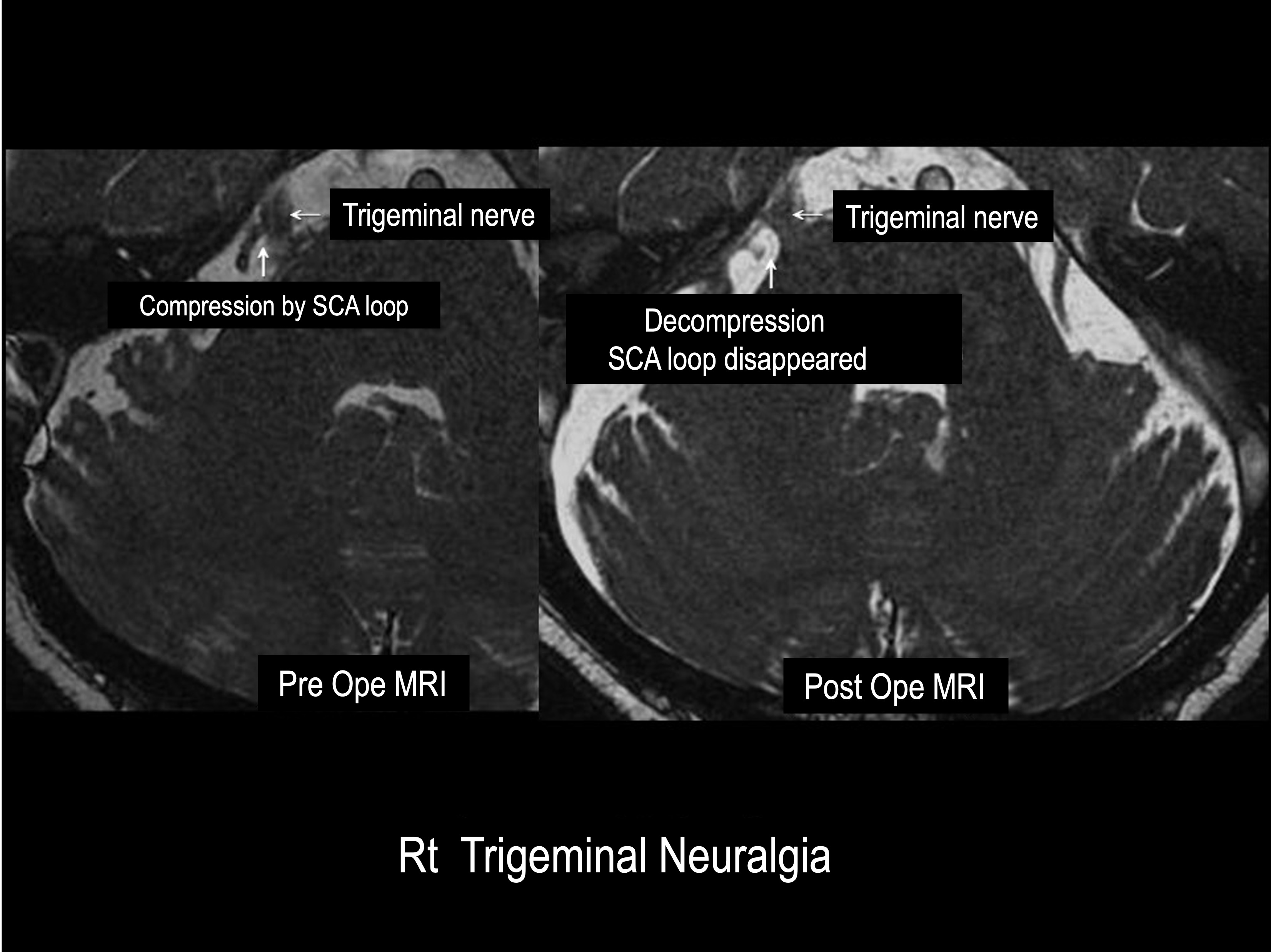
For patients seeking definitive treatment, microvascular decompression (MVD), also known as Jannetta's procedure, remains the most effective curative option. The procedure relieves the vascular compression of the trigeminal nerve by repositioning the offending blood vessel away from the nerve.
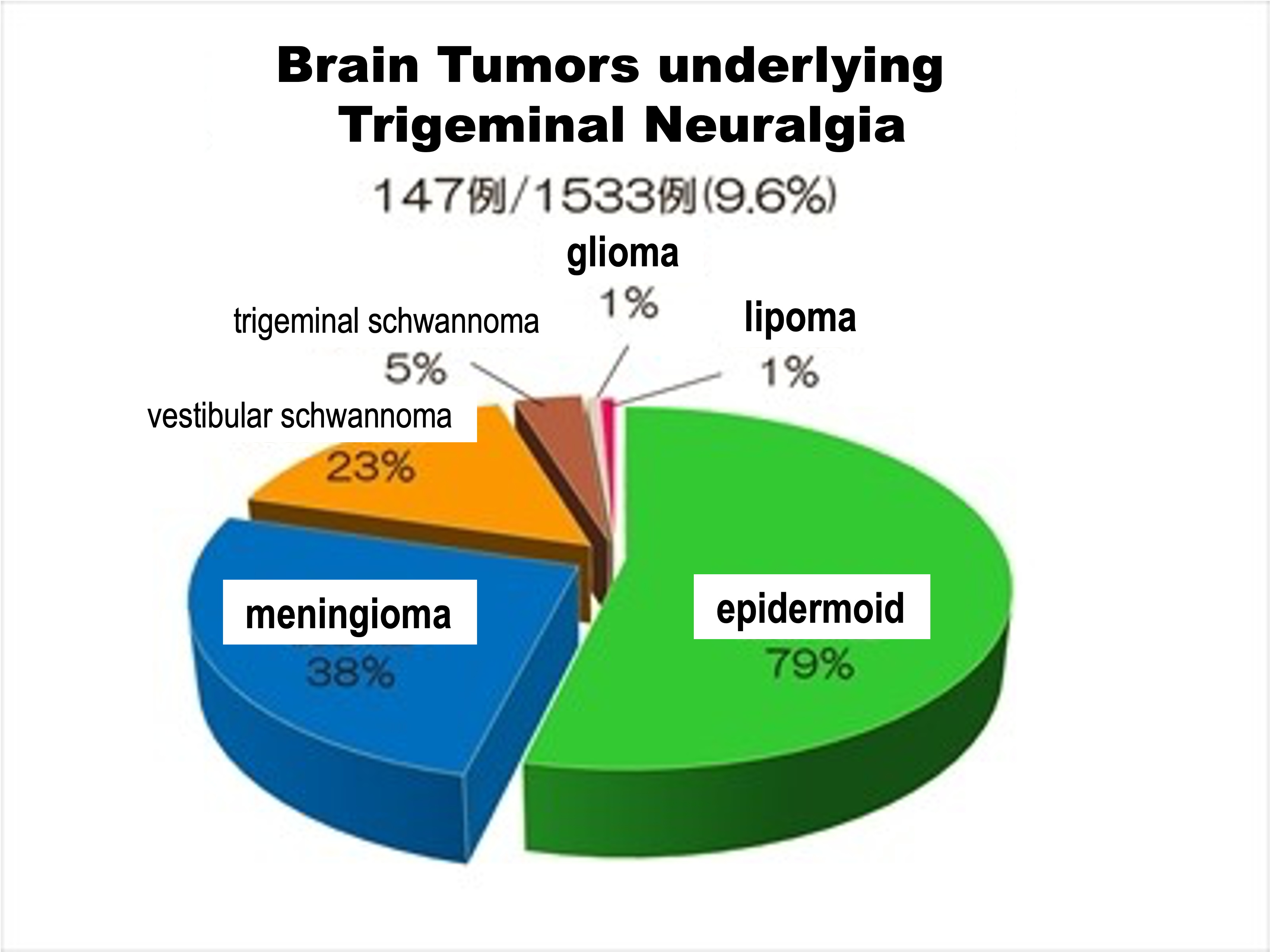
Importantly, studies have reported that approximately one in ten patients diagnosed with trigeminal neuralgia may be found to have an underlying brain tumor. For this reason, patients with trigeminal neuralgia are strongly advised to undergo appropriate neuroimaging studies,
including magnetic resonance imaging (MRI) of the brain, at least once during their evaluation.
In September 2025, the Comprehensive Center for Trigeminal Neuralgia and Hemifacial Spasm was established at Showa University Hospital.
Through close collaboration with the Dental Hospital and related medical specialties, the Center leverages the strengths of a comprehensive academic medical institution to provide multidisciplinary care that is difficult to find elsewhere. Our goal is to deliver coordinated, patient-centered evaluation and treatment for individuals suffering from trigeminal neuralgia, hemifacial spasm, and related cranial nerve disorders.
Diagnostic Imaging
Accurate diagnosis of hemifacial spasm and trigeminal neuralgia requires high-resolution magnetic resonance imaging (MRI).Particularly useful is a specialized MRI technique known as Constructive Interference in Steady State (CISS), which acquires extremely thin-slice images and provides excellent visualization of the cranial nerves and surrounding blood vessels. This imaging method allows detailed assessment of neurovascular relationships and helps identify vascular compression of the affected nerve.
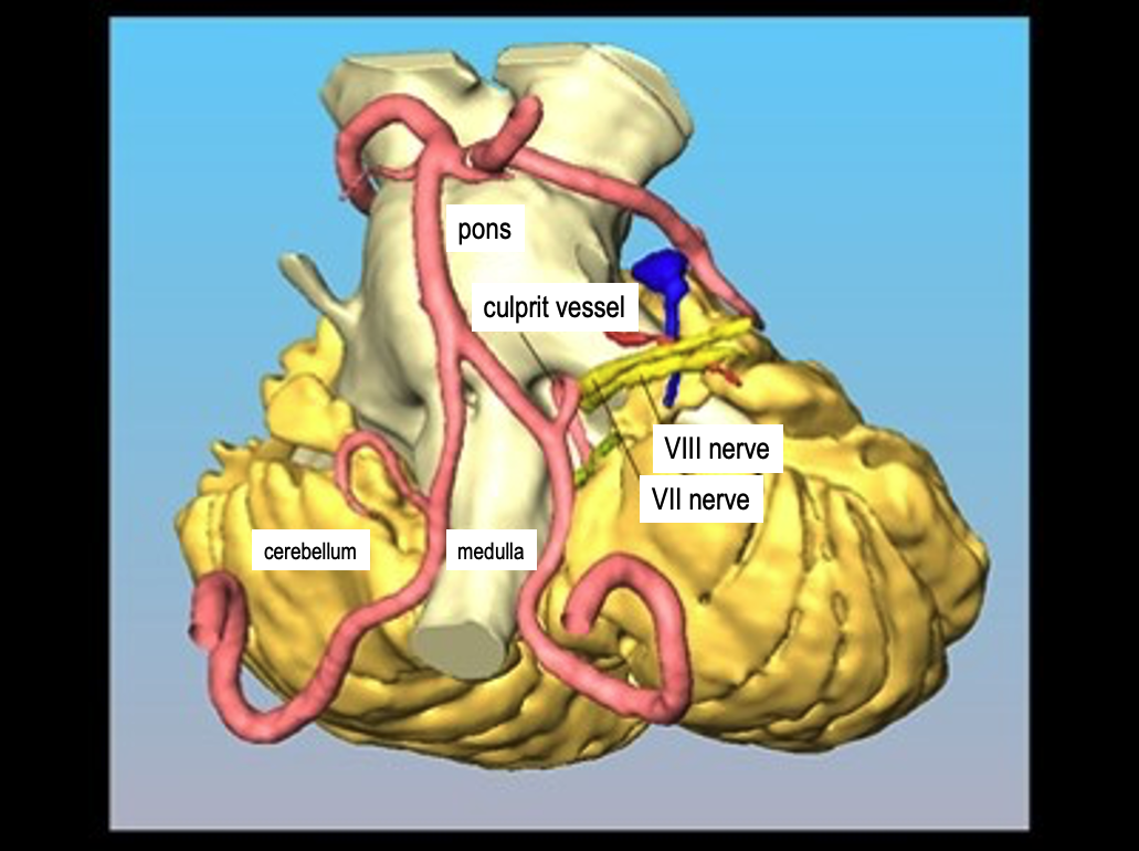
By combining high-resolution MRI with sophisticated 3D image analysis, we are able to achieve a more accurate diagnosis and detailed surgical planning. This contributes to improved surgical safety, greater operative precision, and higher rates of successful treatment.
Surgical Treatment at Our Institution
The Supine No-Retractor Method for Microvascular Decompression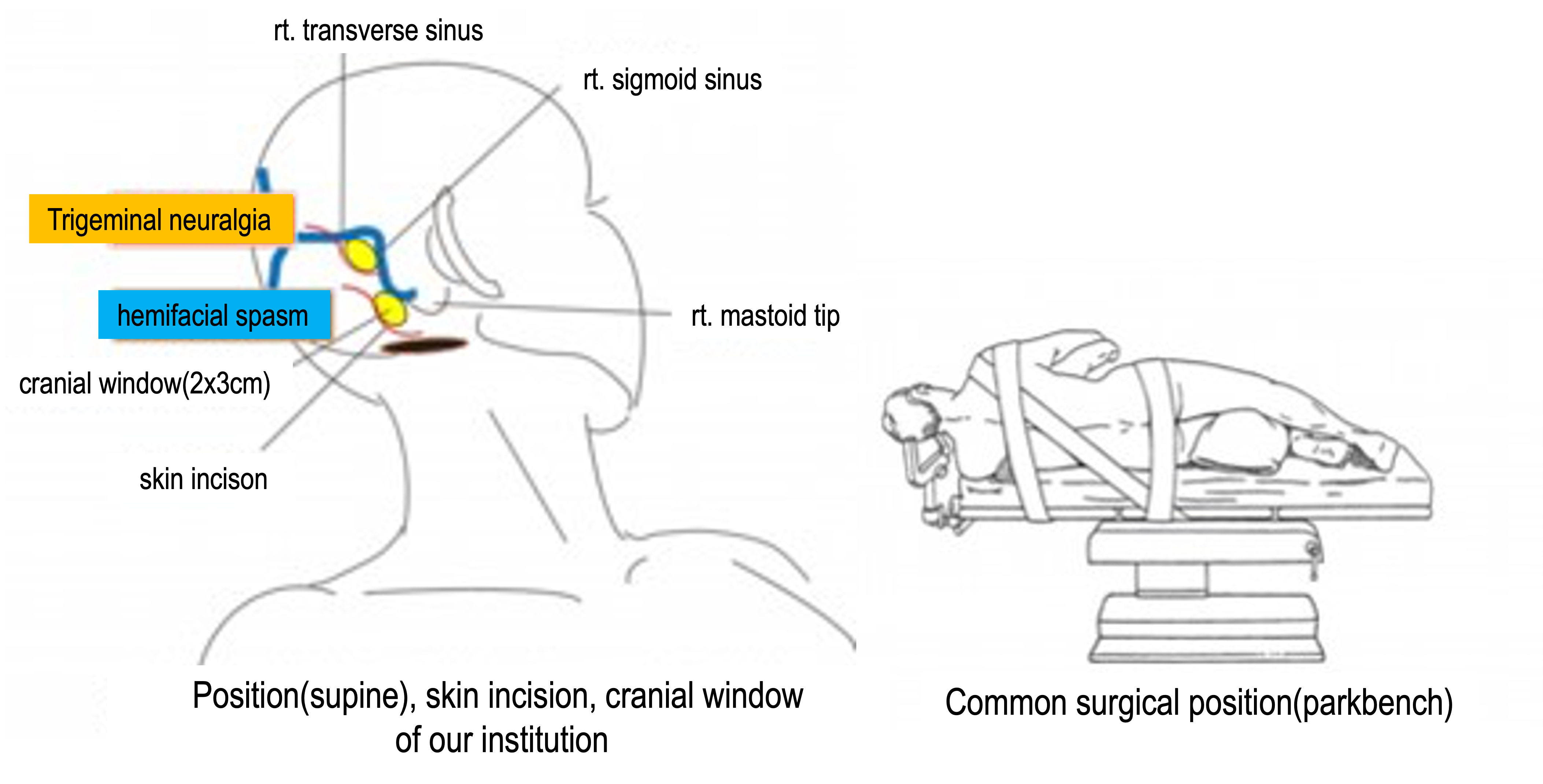
However, repositioning patients into the lateral or prone position after anesthesia can be complex and carries inherent risks. These positions may also place excessive pressure on certain parts of the body, requiring meticulous intraoperative management. Furthermore, excessive cerebellar retraction may increase the risk of complications, including injury to the auditory nerve or cerebellum, potentially resulting in hearing impairment or balance disturbances.
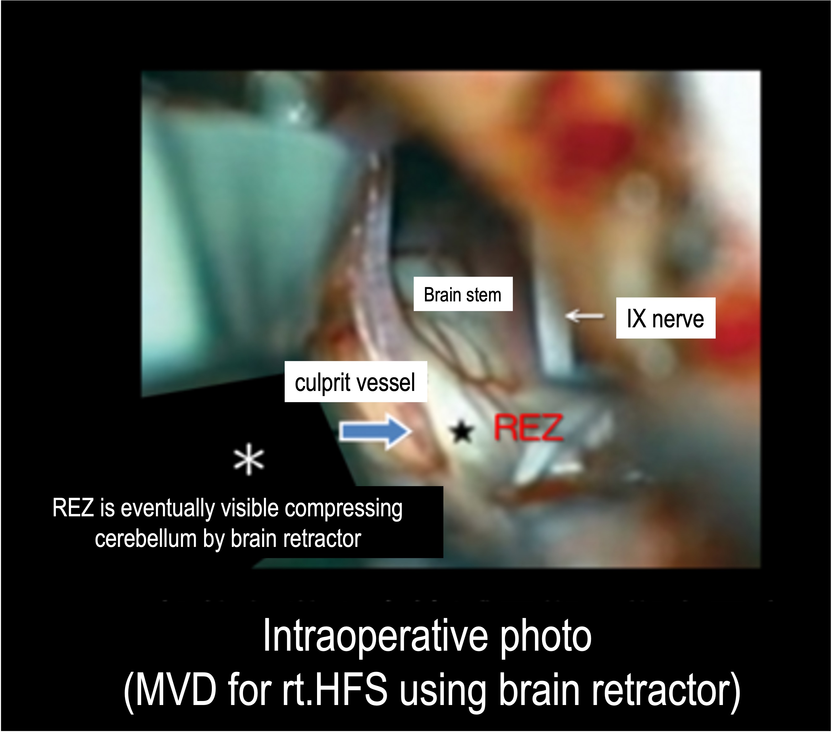
At our institution, we perform MVD with patients lying in the supine position (on their back), the same natural position used during normal sleep. Although this approach requires advanced microsurgical expertise, it takes advantage of the natural downward movement of the cerebellum under its own weight, allowing access to the REZ without the use of brain retractors.
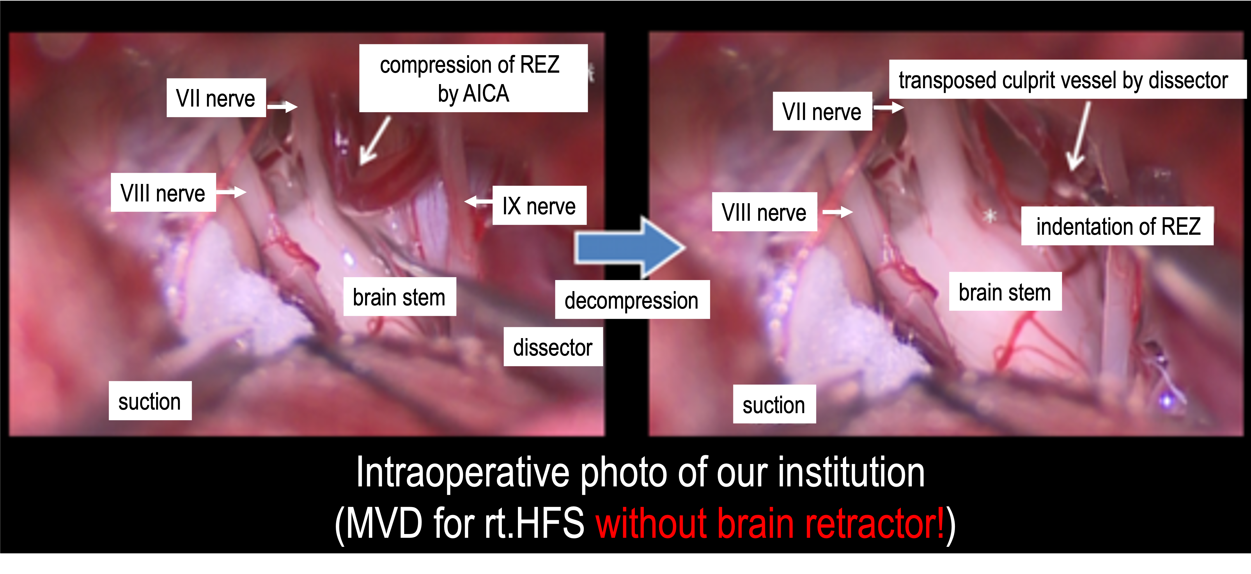
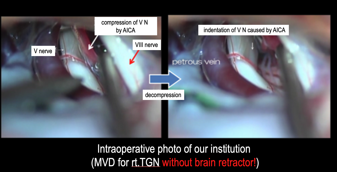
At our institution, all surgical procedures for hemifacial spasm and trigeminal neuralgia are personally performed and supervised by Professor Shimizu, who assumes full responsibility for each patient's care. With extensive experience in microvascular decompression and skull base microsurgery, Professor Shimizu is committed to providing safe, effective, and patient-centered treatment.
We strive to achieve not only excellent surgical outcomes but also a faster and more comfortable postoperative recovery for our patients.
Surgical Outcomes
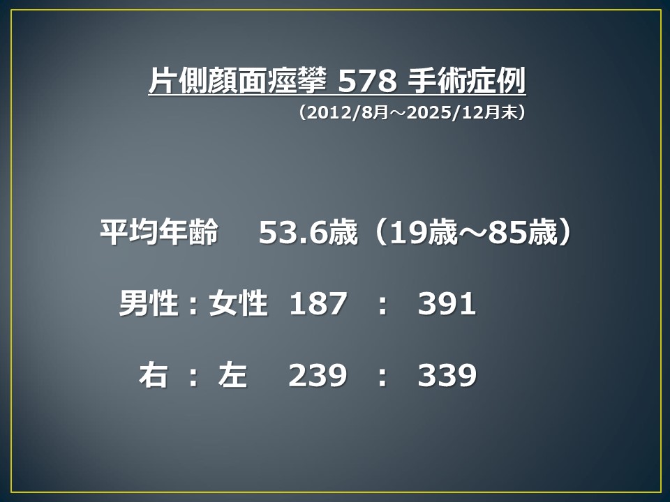
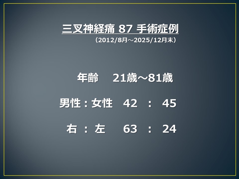
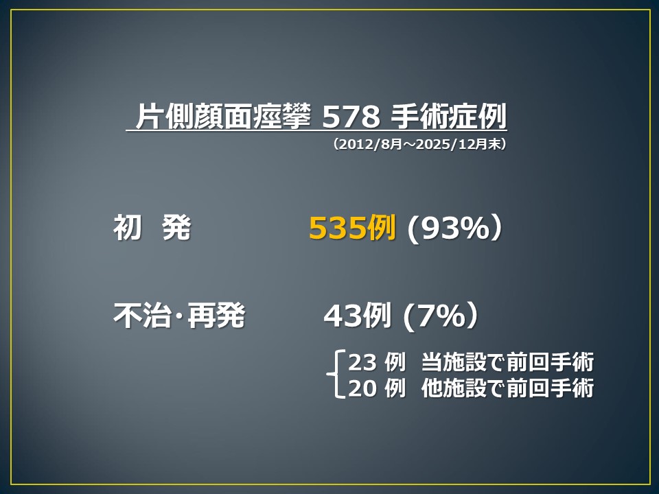
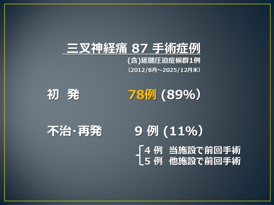
Since Professor Shimizu joined our institution, a total of 578 patients with hemifacial spasm and 87 patients with trigeminal neuralgia have undergone surgical treatment between August 2012 and December 2025.Because the number of trigeminal neuralgia cases is relatively small, the following analysis focuses on patients with hemifacial spasm.
Patient Characteristics
- Total number of cases: 578
- Male: 187 patients
- Female: 391 patients
- Age range: 19 to 85 years
- Mean age: 53.6 years
- Right-sided hemifacial spasm: 239 patients
- Left-sided hemifacial spasm: 339 patients
Surgical Results
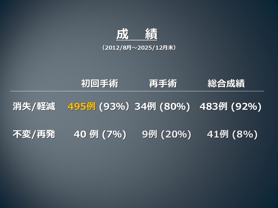
Of the 578 patients treated surgically:- Complete resolution or satisfactory improvement of spasms: 483 patients (92%)
- Persistent symptoms or recurrence: 41 patients (8%)
These results include patients who underwent reoperation for recurrent symptoms.
Complications
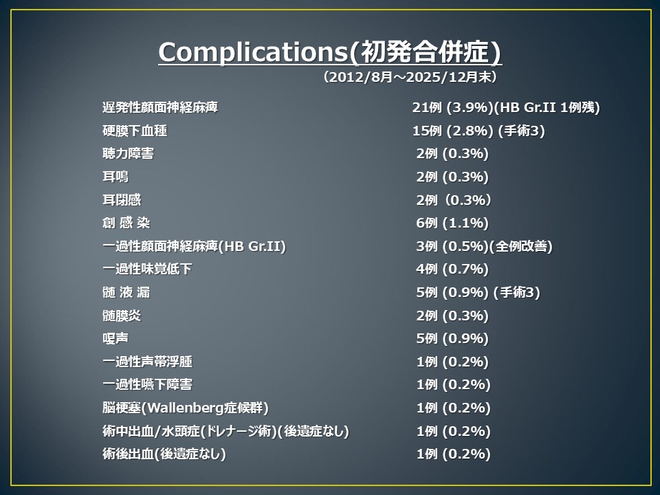
The rates of surgical complications were low:- Hearing deterioration: 2 cases (0.4%)
- Delayed facial nerve palsy: 21 cases (3.9%)
Among patients who developed delayed facial nerve palsy, all but one made a complete recovery. Only a single patient had residual facial weakness.
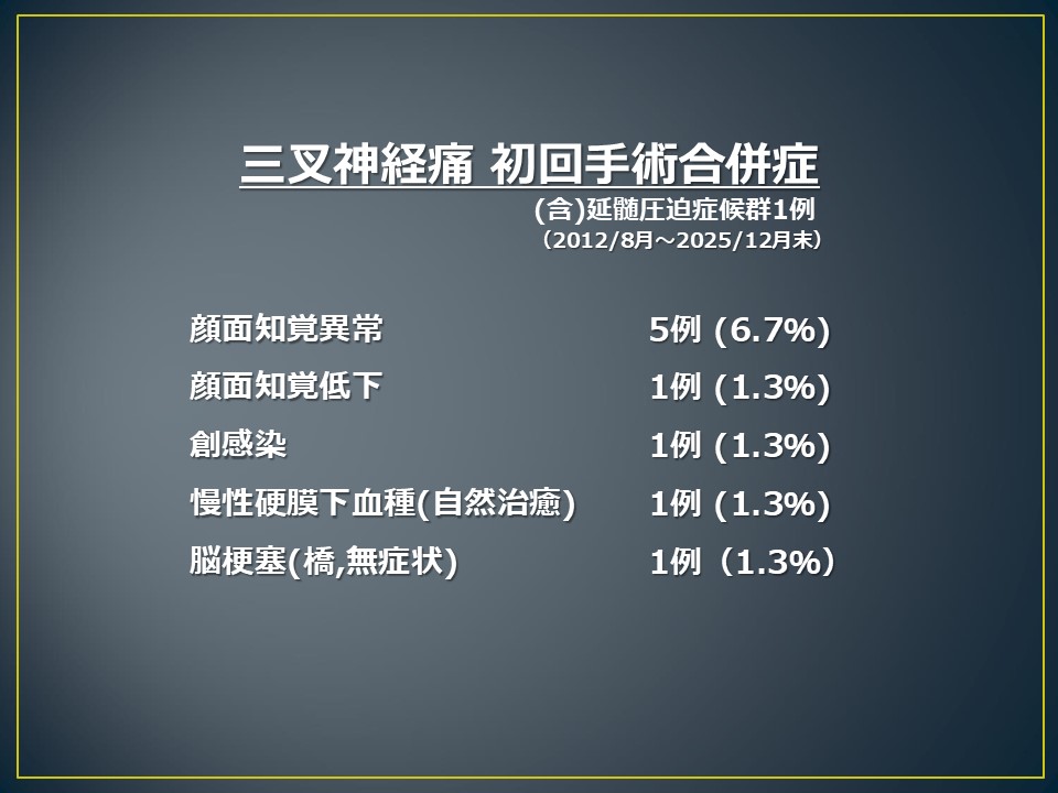
Additional complications are summarized in the accompanying table. Notably, no patient, including those who underwent repeat surgery, experienced a severe permanent neurological deficit or disabling complication.
Our Commitment
These outcomes reflect our ongoing commitment to performing microvascular decompression using the Supine No-Retractor Method, which is designed to minimize surgical trauma while maintaining a high rate of symptom relief. Through meticulous microsurgical technique and careful patient selection, we strive to provide both excellent long-term outcomes and a safe, comfortable recovery for every patient.Reoperation for Persistent or Recurrent Hemifacial Spasm
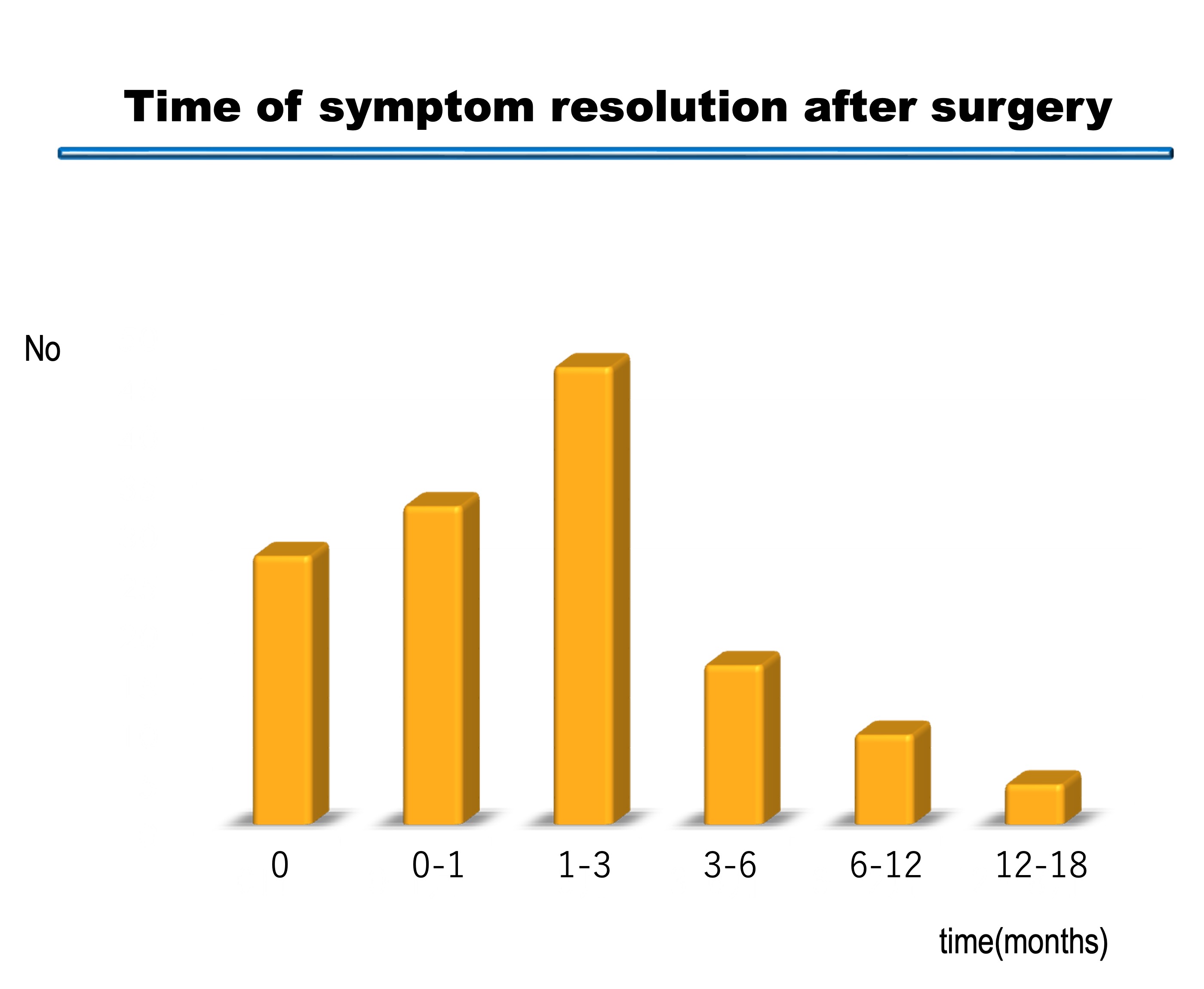
In procedures that cause injury to the facial nerve, patients may develop mild facial weakness immediately after surgery, and the facial spasm may appear to have disappeared right away. In contrast, when surgery is performed without causing damage to the facial nerve, some degree of spasm may remain immediately after the operation. Over time, however, the abnormal hyperactivity of the nerve gradually subsides, leading to progressive improvement and eventual resolution of the spasm.At our institution, it is common for hemifacial spasms to disappear between one and three months after surgery, suggesting that symptom relief is achieved without significant injury to the facial nerve. In some cases, complete resolution has occurred as late as two years after surgery. For this reason, even after an optimal operation, we generally recommend a follow-up period of at least one year before concluding that a case is persistent or recurrent.
Active Recommendation of Reoperation
Even when a patient is ultimately judged to have persistent or recurrent disease, we actively consider and recommend reoperation whenever appropriate.
Several factors may account for persistent or recurrent symptoms. A blood vessel that was not responsible at the time of the initial surgery may later come into contact with the nerve, or multiple offending vessels may have been present from the outset. As is well known, hemifacial spasm and trigeminal neuralgia are typically caused by vascular compression of the nerve near the root exit zone (REZ). Careful preoperative imaging is used to identify the offending vessel, which is then repositioned during surgery.
However, a subset of patients have atypical forms of neurovascular compression, in which the compression occurs at locations other than the REZ. In addition, when large arteries such as the vertebral artery or basilar arteryare involved, the risk of postoperative recurrence may be higher.
Although our institution has achieved a high cure rate following initial surgery, a small number of patients unfortunately do not obtain complete relief. For these patients, reoperation is performed with the goal of identifying and mobilizing any vessels that may still be exerting pressure on the nerve.
Outcomes of Reoperation
In recent years, the medical literature has increasingly supported a proactive approach to reoperation in carefully selected patients. Reports indicate that repeat surgery performed by experienced specialists can achieve successful outcomes even in atypical and treatment-resistant cases.At our institution, the outcomes of reoperation have been encouraging:
- Hemifacial spasm: approximately 80% cure rate
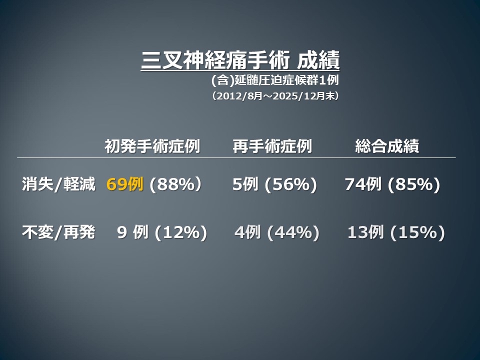
- Trigeminal neuralgia: approximately 56% cure rate
To date, no new major complications have been observed following reoperationat our center.
Our experience has also shown that some patients referred from other institutions had persistent symptoms because the underlying neurovascular compression was not adequately addressed during the initial procedure. In such cases, specialized reassessment and revision surgery may provide an opportunity for meaningful improvement.
Do Not Lose Hope
If you have experienced persistent symptoms or recurrence after your first operation, please do not lose hope. We encourage you to seek a consultation. Even in patients who have had an unfavorable outcome after initial surgery, further evaluation by an experienced microvascular decompression team may reveal treatment options that can still lead to successful relief of symptoms.For more information, please contact the Comprehensive Center for Trigeminal Neuralgia and Hemifacial Spasm.
Attending Neurosurgeon
Professor Katsuyoshi Shimizu, MD, PhDProfessor of Neurosurgery
Director, Comprehensive Center for Trigeminal Neuralgia and Hemifacial Spasm
Showa University Hospital
Please do not hesitate to contact us for consultation regarding hemifacial spasm, trigeminal neuralgia, glossopharyngeal neuralgia, or revision surgery following previous treatment.
Outpatient Consultation
- Professor Shimizu's outpatient clinic is held on Thursdays. Please feel free to schedule a consultation.
- Professor Shimizu may occasionally be unavailable due to surgery, academic meetings, or other professional commitments. We would appreciate it if you could call in advance to confirm his availability before your visit.
Medical Liaison Office: +81-3-3784-8400 e-mail: neurosurg@cnt.showa-u.ac.jp
Featured Articles and Physician Profiles
Professor Katsuyoshi Shimizu has been featured in a number of medical information resources and physician directories:- Brain Disease Web
Professor Shimizu Featured on Brain Disease Web - Doctor's Guide
Professor Shimizu Featured in Doctor's Guide - Medical Note
Professor Shimizu Featured on Medical Note - Neurosurgical Specialists Directory
Professor Shimizu Featured in the Neurosurgical Specialists Directory - CocoKaraDa Hospital Search
Professor Shimizu Featured on CocoKaraDa