- SHOWA Medical University Hospital
- Benign Brain Tumors, Skull Base Tumors, and Orbital Tumors
- Japanese
- English
Benign Brain Tumors, Skull Base Tumors, and Orbital Tumors
Introduction
Brain tumors are estimated to occur in approximately one person per 10,000. Among these, benign brain tumors are tumors that do not metastasize to other parts of the body, grow slowly, and are usually well demarcated from the surrounding brain tissue. They account for approximately 40-45% of all brain tumors. Common examples include meningiomas, schwannomas, and pituitary adenomas. Complete surgical removal can often provide a cure.Symptoms may include headaches, dizziness, seizures, and cognitive impairment. Depending on the location of the tumor, a wide variety of neurological symptoms can occur. With the widespread use of CT and MRI, an increasing number of tumors are being discovered incidentally during examinations performed for other reasons.
Even benign tumors can present significant challenges when they arise in or extend into the skull base, the region at the base of the skull where critical blood vessels and numerous cranial nerves are concentrated. Tumors in this area frequently involve these vital structures, making treatment more complex and potentially affecting long-term outcomes.
Less invasive Tumor Surgery Performed in the Supine Position Without Brain Retractors
Dr. Katsuyoshi Shimizu specializes in delicate microsurgical procedures, including keyhole surgery and the treatment of skull base tumors. His expertise has been recognized by several independent medical information resources, including:- Doctor's Guide (Jiji Press Publishing)
http://dr-guide.net/www/ - List of Distinguished Neurosurgeons
http://doctor-cancer.com/cranialnerves-dr.html - Brain Disease Web: Distinguished Neurosurgeons
Dr. Shimizu is particularly known for minimally invasive surgical techniques that prioritize the preservation of normal brain tissue. By performing surgery in the supine position and avoiding the use of brain retractors whenever possible, he strives to minimize surgical stress on the brain and enhance patient safety and recovery.
Through meticulous microsurgical techniques and extensive experience in skull base surgery, our team aims to achieve maximal tumor removal while preserving neurological function and quality of life.


Media Recognition and Surgical Expertise
Dr. Katsuyoshi Shimizu was featured as the treating neurosurgeon and operating surgeon of a well-known actress who successfully recovered from hemifacial spasm following surgery. Her story was broadcast on Fuji TV's FNN Super News in 2010 and TBS's Hiruobi! in 2011. Dr. Shimizu also appeared on the nationally televised health program "Takeshi's Health Entertainment! The Family Doctor" in 2012.Since 1985, Dr. Shimizu has performed microvascular decompression (MVD), the definitive surgical treatment for hemifacial spasm and trigeminal neuralgia, using a unique technique in which patients are positioned supine (lying on their back). He has published this method in the scientific literature and established it as a distinctive surgical approach.
This technique is particularly patient-friendly because it utilizes the body's most natural position, the supine position, and avoids the use of a brain retractor, a device commonly employed during neurosurgical procedures to retract brain tissue. By minimizing manipulation of the brain, the procedure aims to reduce surgical stress and improve patient safety.
Approximately one in ten patients with trigeminal neuralgia is found to harbor an underlying brain tumor, highlighting the importance of careful diagnostic evaluation. As a result, Dr. Shimizu has also accumulated extensive experience in the surgical treatment of brain tumors.
The supine, retractor-free surgical approach has been successfully applied not only to microvascular decompression procedures but also to the treatment of vestibular schwannomas (acoustic neuromas) and other skull base tumors. Dr. Shimizu is widely recognized for his expertise in these highly delicate microsurgical procedures, which require meticulous technique to preserve critical nerves and blood vessels while achieving optimal treatment outcomes.
Meningioma
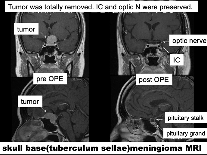
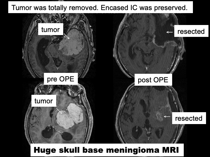
Depending on their location, meningiomas are classified into several types, including:
- Convexity meningiomas
- Parasagittal meningiomas
- Sphenoid wing meningiomas
- Tentorial meningiomas
- Falcine meningiomas
- Posterior fossa meningiomas
Convexity meningiomas, which develop on the surface of the brain, often grow slowly and may produce few symptoms. As a result, some tumors are not detected until they become quite large. In certain cases, patients who have been treated for dementia because of behavioral or cognitive symptoms are ultimately found to have a giant frontal lobe meningioma.
Depending on the tumor location, a variety of neurological deficits may occur, including:
- Visual impairment
- Facial sensory disturbances
- Hearing loss
- Difficulty swallowing
- Balance problems
- Weakness or numbness
Tumors such as sphenoid wing meningiomas, cerebellopontine angle (CPA) meningiomas, and clival meningiomas are classified as skull base tumors. Because these tumors arise in regions densely populated by critical cranial nerves and blood vessels, their treatment is often particularly challenging.
Similarly, parasagittal meningiomas frequently invade the superior sagittal sinus, a major venous channel of the brain, as they enlarge. Such involvement can significantly increase the complexity of surgical treatment.
Treatment
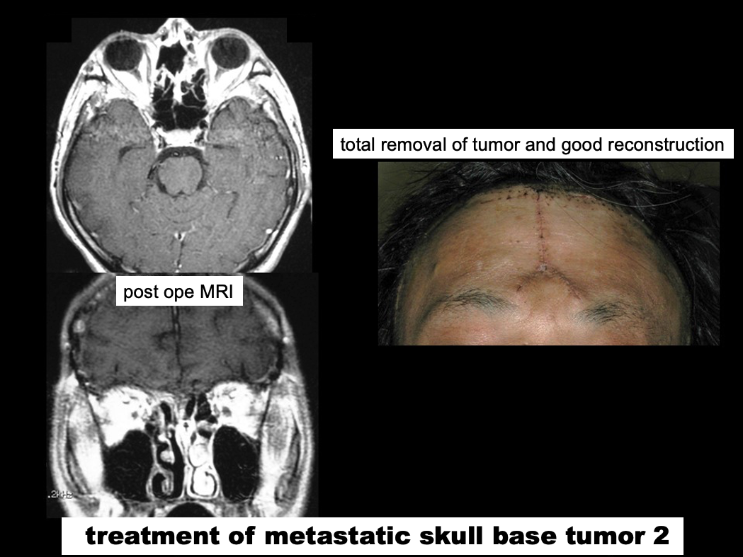
The primary treatment for meningiomas is surgical removal.Small convexity meningiomas can often be removed relatively easily. However, more complex tumors, such as skull base meningiomas and giant meningiomas, frequently encase or adhere to important nerves and blood vessels. In these cases, successful treatment requires an experienced surgeon with specialized expertise in skull base microsurgery.
When complete removal is not possible because of involvement of critical neurovascular structures, or in the uncommon event that pathological examination reveals more aggressive tumor features associated with a higher risk of recurrence, stereotactic radiosurgerysuch as Gamma Knife or CyberKnife may be recommended following surgery.
Our Approach
At our institution, specialists with extensive experience in the management of meningiomas carefully evaluate all available treatment options, including:- Microsurgical resection
- Stereotactic radiosurgery
- Radiation therapy
- Observation with serial imaging
Rather than applying a uniform treatment strategy, we provide personalized, patient-centered care, developing an individualized treatment plan tailored to each patient's tumor characteristics, symptoms, age, overall health, and personal preferences. Our goal is to achieve the best possible tumor control while preserving neurological function and quality of life.
Schwannomas (Vestibular Schwannomas / Acoustic Neuromas)
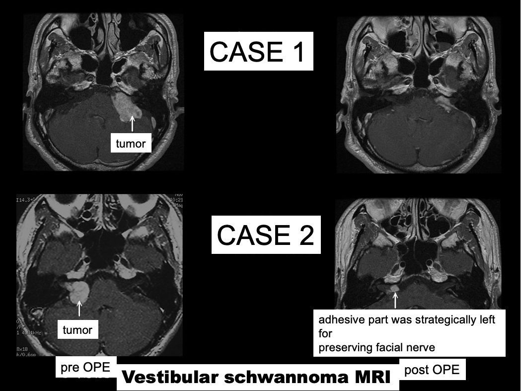
Schwannomas account for approximately 10% of all primary brain tumors. They most commonly arise from the vestibular nerve (often referred to as an acoustic or vestibular schwannoma) and the trigeminal nerve.
Symptoms
Symptoms depend on the nerve from which the tumor originates and on the neighboring nerves or structures affected by the tumor.Vestibular schwannomas may cause:
- Hearing loss
- Tinnitus (ringing in the ear)
- Dizziness or imbalance
Trigeminal schwannomas may cause:
- Facial numbness or sensory disturbances
- Facial pain
- Double vision (due to involvement of nerves controlling eye movements)
Treatment
As with meningiomas, the primary treatment for schwannomas is surgical removal.As a schwannoma enlarges, the original nerve fibers are gradually compressed and stretched over the surface of the tumor, becoming extremely thin, almost like a sheet of paper. These compressed nerve fibers are highly vulnerable to surgical manipulation.
For example:
- In vestibular schwannomas, injury to the nerve may result in hearing loss.
- In trigeminal schwannomas, injury may cause facial numbness or sensory deficits.
Successful schwannoma surgery therefore requires a specialized microsurgical technique. Rather than simply removing the entire mass en bloc, the surgeon carefully removes the tumor while preserving the delicate outer layer, or capsule, that contains the flattened nerve fibers adherent to the tumor surface.
Vestibular Schwannoma: A Particularly Challenging Tumor
Among all schwannomas, vestibular schwannomas are considered the most technically demanding.These tumors arise in the cerebellopontine angle (CPA), a complex region where numerous cranial nerves and major blood vessels are densely concentrated. The facial nerve, which controls facial movement, is often tightly adherent to the tumor surface.
If the facial nerve is damaged during tumor removal, patients may develop facial weakness or facial paralysis following surgery. Consequently, meticulous microsurgical dissection is essential to preserve facial nerve function.
Special attention is required in patients who retain useful hearing before surgery. In selected cases, particularly when hearing remains good and the tumor is not growing significantly, careful observation rather than immediate surgery may be the most appropriate management strategy.
Adjuvant Radiation Therapy
In some cases, complete tumor removal may not be possible because of strong adhesion to critical nerves or blood vessels. In other rare situations, pathological examination may reveal more aggressive features associated with a higher risk of recurrence.Under such circumstances, stereotactic radiosurgery, including:
- Gamma Knife®
- CyberKnife®
may be recommended following surgery, similar to the treatment strategy employed for certain meningiomas.
Our Approach
At our institution, patients with schwannomas are evaluated by specialists with extensive experience in skull base and cranial nerve surgery. We consider all available treatment options, including:- Microsurgical resection
- Stereotactic radiosurgery
- Observation with serial MRI follow-up
Rather than applying a uniform treatment strategy, we provide personalized, tailor-made care, developing an individualized treatment plan based on each patient's tumor characteristics, symptoms, neurological function, age, overall health, and personal preferences. Our goal is to achieve optimal tumor control while preserving neurological function and maintaining the highest possible quality of life.
Skull Base Tumors
The skull base refers to the bottom portion of the skull that supports and contains the brain. Tumors arising in this region are collectively known as skull base tumors, a term that has become widely used in modern neurosurgery.A variety of tumors may occur at the skull base, including benign lesions such as:
- Meningiomas (e.g., sphenoid wing, cerebellopontine angle, and clival meningiomas)
- Schwannomas (e.g., vestibular, trigeminal, and jugular foramen schwannomas)
- Giant pituitary adenomas
In addition, certain malignant tumors originating in the fields of otolaryngology (ENT) and ophthalmology may also involve the skull base.
Why Are Skull Base Tumors Challenging?
Beneath the skull base lie many of the structures that make up the face, including the eyes, ears, nose, mouth, and throat. The nerves that control these organs originate in the brain and pass through the skull base before reaching their respective targets.Furthermore, all major intracranial arteries converge at the skull base to form the Circle of Willis, from which the blood supply to the entire brain is distributed.
As a result, even benign skull base tumors frequently involve critical blood vessels and cranial nerves. Treatment therefore carries a significant risk of neurological complications, including:
- Loss of smell
- Visual impairment
- Abnormal eye movements or double vision
- Hearing loss
- Difficulty swallowing
- Speech and articulation problems
For this reason, skull base tumors have traditionally been regarded as one of the most challenging groups of neurosurgical diseases. In the past, this region was often referred to as a "no man's land" because of the difficulty and risks associated with surgical treatment. However, advances in microsurgical techniques, intraoperative monitoring, imaging technology, and surgical instruments have made treatment significantly safer and more effective than ever before.
Our Surgical Approach
At our institution, skull base tumor surgery is performed by highly experienced surgeons specializing in skull base microsurgery.To maximize safety and precision, we utilize advanced technologies including:
- Image-guided surgical navigation systems
- Auditory Brainstem Response (ABR) monitoring
- Comprehensive intraoperative neurophysiological monitoring
- High-resolution microsurgical techniques
Particular attention is also paid to patient positioning and head orientation. Whenever possible, we employ techniques that minimize or eliminate the need for brain retractors, thereby reducing pressure on normal brain tissue and helping to preserve neurological function.
Function-Preserving, Multidisciplinary Treatment
Our guiding principle is to achieve effective tumor control while avoiding new neurological deficits whenever possible.When a tumor is densely adherent to major blood vessels, cranial nerves, or the brainstem, complete removal may not always be in the patient's best interest. In such situations, we may intentionally leave a thin layer of tumor tissue to preserve critical neurological structures and subsequently treat any residual tumor with adjunctive therapies such as:
- Gamma Knife® radiosurgery
- CyberKnife® radiosurgery
- Other forms of focused radiation therapy
This individualized, multidisciplinary strategy allows us to maximize both tumor control and functional preservation.
Collaborative Care
In addition to tumors originating within the brain, we also treat skull base tumors arising from neighboring regions managed by other specialties.For these complex cases, we work closely with specialists in:
- Otolaryngology (ENT)
- Ophthalmology
- Plastic and Reconstructive Surgery
Through this multidisciplinary collaboration, we provide comprehensive, patient-centered care tailored to the specific characteristics of each tumor and the individual needs of each patient. Our ultimate goal is to achieve the best possible oncological outcome while preserving neurological function and quality of life.
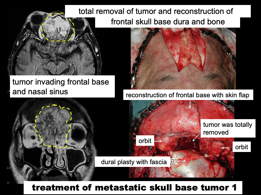
Orbital Tumors
The orbit is the bony cavity in the front of the skull that contains and protects the eyeball. Because the orbit contains a variety of tissues, including nerves, blood vessels, fat, and exocrine glands, a wide range of tumors can arise within this confined space, although orbital tumors are relatively uncommon.Reports from Japan indicate that more than half of orbital tumors are lymphoproliferative lesions, with malignant lymphoma accounting for approximately 15-30% of cases. Other relatively common tumors include cavernous hemangiomasand pleomorphic adenomas of the lacrimal gland, each representing approximately 10% of cases. Less common lesions, each accounting for fewer than 5% of cases, include dermoid cysts, meningiomas, and adenoid cystic carcinomas, among others.
Symptoms
The symptoms of orbital tumors vary depending on the tumor's location, size, and relationship to surrounding structures. Possible symptoms include:- Visual impairment
- Visual field defects
- Restriction of eye movements
- Double vision
- Protrusion of the eye (proptosis)
- Eye pain
If surgical treatment is not performed at an appropriate stage, some of these symptoms may become permanent. Therefore, prompt evaluation and treatment by specialists are strongly recommended whenever an orbital tumor is suspected.
Diagnosis and Preoperative Evaluation
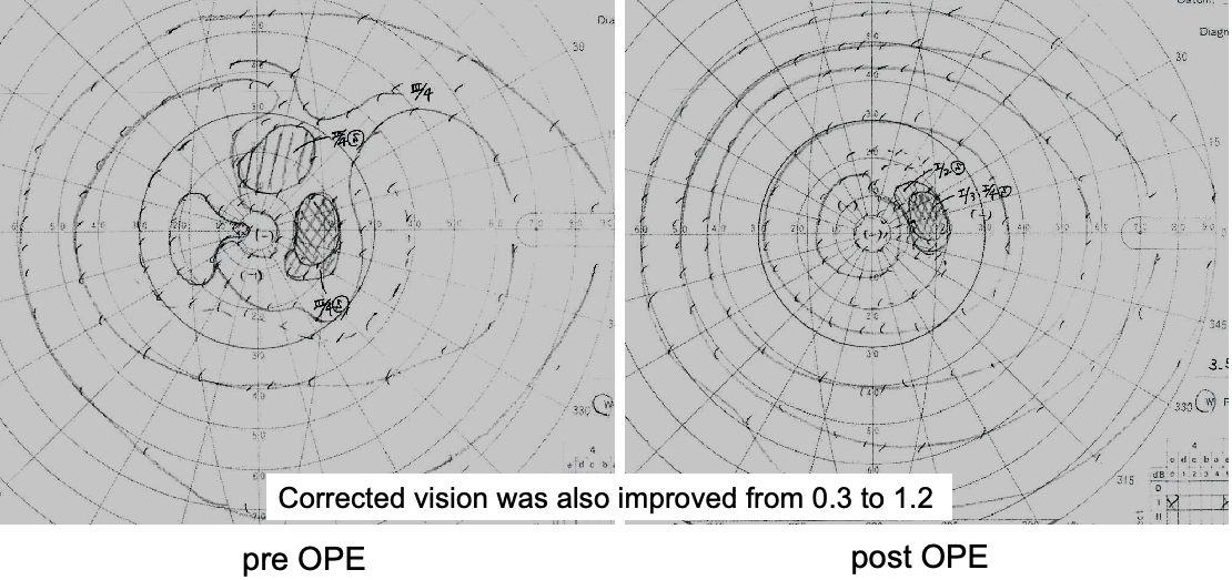
Computed tomography (CT) and magnetic resonance imaging (MRI) are indispensable for the diagnosis and characterization of orbital tumors. Detailed preoperative assessment is essential to determine the exact location and extent of the lesion and to plan the safest and most effective surgical approach.Our team works closely with ophthalmologists both before and after surgery to evaluate visual function, including visual acuity, visual fields, and ocular motility, ensuring comprehensive care throughout the treatment process.
Surgical Treatment
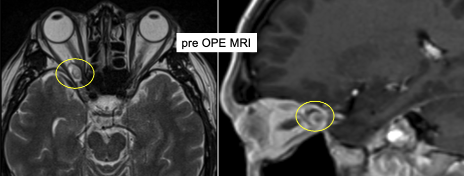
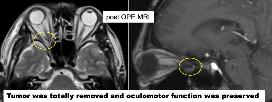
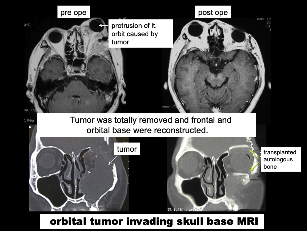
Even relatively small tumors located at the orbital apex, the deepest portion of the orbit, can cause significant symptoms because of compression of the optic nerve and other critical neurovascular structures.For such lesions, as well as many other orbital tumors, we believe that a transcranial approach often provides the safest and most reliable access, allowing precise tumor removal while minimizing the risk of damage to the optic nerve and surrounding structures. Accordingly, this approach is routinely employed at our institution when indicated.
In one representative case, although the patient was referred to our center some time after symptom onset, surgery resulted in an excellent outcome, with visual acuity and visual fields recovering to near-normal levels after treatment. The patient was able to make a rapid postoperative recovery and was discharged shortly thereafter.
Our Approach
At our institution, orbital tumor treatment is provided through close collaboration between neurosurgery and ophthalmology, with individualized treatment plans tailored to each patient's condition. Our goal is to preserve vision and ocular function while achieving maximal safe tumor removal.If you have been diagnosed with an orbital tumor or are experiencing unexplained visual symptoms, we encourage you to consult our specialists for a thorough evaluation and expert treatment.
Final Message
Medicine should be grounded in science, and in recent years, with the growing emphasis on evidence-based medicine (EBM), medical practice has become increasingly standardized and universally applicable. Nevertheless, even within university hospitals that are expected to provide the most advanced medical care, there remains a tendency to focus on the disease itself rather than on the individual patient.In reality, however, medical care is not a science that produces exactly the same outcome regardless of when, where, or by whom it is delivered. The very existence of the term “master physician” reflects this truth. While medicine is founded on scientific knowledge, healthcare is ultimately an art practiced by one human being for another.
Every patient is unique, and the medical care required by each individual differs accordingly. Truly ideal healthcare can only be achieved by combining treatments based on the latest scientific evidence with the refined judgment, experience, and skill that come from years of clinical practice. It is this balance of science and art that enables physicians to provide the best possible care for each patient.
Our goal is to be not only scientists who are thoroughly familiar with the latest advances in medicine, but also artists of medicine who understand each patient's concerns, suffering, and clinical condition. By listening carefully and treating every individual with compassion and expertise, we strive to provide the highest standard of personalized care to every patient entrusted to us.
Selected References
- Yamaguchi S, et al. Shinkei Chiryo (Neurological Therapeutics). 2010;27:107-132.
- Shimizu K, et al. Lateral basal approach with a supine, no-retractor method for microvascular decompression for hemifacial spasm. Acta Neurochirurgica. 2015;157(5):803-806.
DOI: 10.1007/s00701-015-2393-9 - Shimizu K, et al. Supine no-retractor method in microvascular decompression for hemifacial spasm: Results of 100 consecutive operations. Journal of Neurological Surgery Part B: Skull Base. 2015;76(3):202-207.
DOI: 10.1055/s-0034-1396660 - Shimizu K, et al. Lateral basal space and interlaminar fatty compartment of the deep cervical fascia in the posterolateral craniocervical junction: An anatomical basis for surgery of the lateral skull base. International Journal of Neurology and Neurotherapy. 2016;3:038.
ISSN: 2378-3001
This version maintains the philosophical and patient-centered message of the original Japanese text while adopting a polished, professional tone appropriate for an academic medical center website and international readership.
Attending Physician
Katsuyoshi Shimizu, MD, PhD
Dr. Katsuyoshi Shimizu specializes in microsurgical treatment of hemifacial spasm and trigeminal neuralgia, as well as skull base tumors, including:- Vestibular schwannomas (acoustic neuromas)
- Pituitary tumors
- Meningiomas
- Other complex skull base lesions
With extensive experience in advanced microsurgical techniques, Dr. Shimizu is particularly recognized for minimally invasive approaches designed to maximize tumor removal and symptom relief while preserving neurological function and quality of life.
Outpatient Clinic
- Clinic Day:Thursday
- Patients are welcome to schedule a consultation and discuss their condition with us.
Please note that Dr. Shimizu may occasionally be unavailable due to surgery, academic meetings, or other professional commitments. We would appreciate it if you could confirm his availability by telephone before your visit.
Medical Liaison Office:+81-3-3784-8400
e-mail:neurosurg@cnt.showa-u.ac.jp
Patients with hemifacial spasm, trigeminal neuralgia, vestibular schwannomas, meningiomas, pituitary tumors, and other skull base disorders are encouraged to contact our center for expert evaluation and individualized treatment planning.